Cnicothamnus lorentzii (Asteraceae): foliar morphology, anatomy and architecture
Keywords:
Anatomy, Cnicothamnus, leaf, stomata, trichomesAbstract
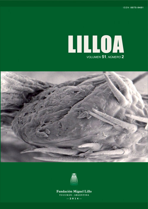
Ruiz, Ana I.; María E. Guantay; María I. Mercado; Graciela I. Ponessa. 2014. “Cnicothamnus lorentzii (Asteraceae): foliar morphology, anatomy and architecture”. Lilloa 51 (2). Cnicothamnus lorentzii Griseb., is a native shrub distributed along the provinces of Tucumán, Salta and Jujuy and in the neighboring country of Bolivia. It is of medicinal interest, since its leaves and stems exhibit antilesmanial and trypanocidal activity. The aim of this work is to describe the morphology, anatomy and leaf architecture of C. lorentzii from Trancas, Tucuman, Argentina to identify characters of diagnostic value. Samples were processed using standards techniques for light and electron microscopy. Cnicothamnus lorentzii has simple leaves; the adaxial and abaxial sur faces are sub-glabrous and pubescent respectively. Glandular and eglandular trichomes were observed. Epidermal cells are polygonal with lobed anticlinal walls. In transverse section is dorsiventral, hypostomatic with con anomocytic, hemibrachy-paracytic, tetracytic and pentacytic. The midrib presents six collateral vascular bundles. The pubescent petiole shows 11 collateral vascular bundles arranged in a semicircle. Venation is pinnate camptodromous-eucamptodromous. Secondary veins straight. Areoles with and without freely ending veinlets. First described the anatomy and architecture and leaf petiole of C. lorentzii. The diagnostic value features are: leaf architecture, types of stomata and trichomes
Downloads
References
Barboza G., Cantero J., Núñez C., Pacciaroni A., Ariza Espinar L. 2009. Medicinal plants: A general review and a phytochemical and ethnopharmacological screening of the native Argentine Flora. Kurtziana 34 (1-2): 7-365.
Bianco C., Kraus T., Vegetti A. 2004. La hoja: Morfología externa y anatomía. Editora Universidad de Río Cuarto y Universidad Nacional del Litoral, Argentina, 199 pp.
D’Ambrogio de Argüeso A. 1986. Manual de Técnicas en Histología Vegetal. Editora Hemisferio Sur S. A., Buenos Aires, 83 pp.
Digilio A., Legname P. 1966. Los árboles indígenas de la provincia de Tucumán. Opera Lilloana 15: 20, 129 pp.
Dilcher D. 1974. Approaches to the identification of angiosperm leaves. The Botanical Review 40 (1): 1-157.
Dimitri M. 1972. Enciclopedia Argentina de Agricultura y Jardinería. Editora Acme‚ S.A.C.I. Buenos Aires, 1028 pp.
Dizeo de Strittmater C. 1973. Nueva técnica de diafanización. Boletín de la Sociedad Argentina de Botánica 15 (1): 126-129.
Ellis B., Daly D., Hickey L., Johnson K., Mitchell J., Wilf P., Wing S. 2009. Manual of leaf architecture. Cornell University Press, USA, 190 pp.
Ehleringer J., Mooney H. 1978. Leaf hairs: effects on physiological activity and adaptative value to a desert shrub. Oecologia 37: 183-200.
Esau K. 2008. Anatomía vegetal. Editora Omega, Barcelona, España, 641 pp.
Fahn A. 1986. Structural and functional properties of trichomes of xeromorphic leaves. Annalen der Botanick (London) 57: 631-637.
Fournet A., Angelo Barrios A., Muñoz V. 1994. Leishmanicidal and trypanocidal activities of Bolivian medicinal plants. Journal of Ethnopharmacology 41: 19-37.
Hickey L. 1974. Clasificación de la arquitectura de las hojas de Dicotiledóneas. Boletín Sociedad Argentina Botánica 16 (1-2): 1-26.
Hickey L. 1979. A revised classification of the architecture of dicotyledonous leaves. En: Metcalfe C., Chalk L. (editores), Anatomy of the Dicotyledons. Volumen I. Second Edition. Clarendon Press, Oxford: 25-39.
Johnson H. 1975. Plant pubescence: an ecological perspective. Botanical Review 41: 233-258.
Karnovsky M. 1965. A formaldehyde glutaraldehyde fixative of high osmolality for use in electron microscopy. Journal of Cell Biology 27:137-138.
Merck E. 1980. Reactivos de coloración para cromatografía en capa fina y en papel. R. F., Alemania, 119 pp.
Metcalfe C., Chalk L. 1950. Anatomy of the Dicotyledons. Editora Clarendon Press, Oxford, pp 1145-1156.
Nógalo A., Molina G., Norry G., Romano S., Lorenz A. 2012. Leishmaniasis cutánea primaria. Dermatología Argentina 18 (3): 228-230.
Novara L., Katinas L., Urtubey E. 1995. Flora del Valle de Lerma. Asteraceae. Tribu 10. Mutisieae Cass. Aportes Botánicos de Salta. Serie Flora 3 (1): 21-23.
Roth I. 1984. Stratification of tropical forest as seen in leaf structure. Editor Junk, University of Minnesota. 521 pp.
Solereder H. 1908. Systematic anatomy of the Dicotyledons. Oxford at the Clarendon Press. 895 pp.
Zuloaga F., Morrone O., Belgrano M. 2008. Catálogo de Plantas Vasculares del Cono Sur (Argentina, Sur de Brasil, Chile, Paraguay y Uruguay). Editora Missouri Botanical Garden, Saint Louis, Missouri, 3348 pp.