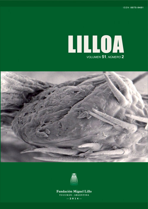
Morphology and anatomy of extrafloral nectaries in three species of Flacourtiaceae (Salicaceae sensu lato)
Keywords:
anatomy, extrafloral nectaries, Flacourtiaceae, morphologyAbstract
Cortadi, Adriana; Lourdes Pulido García; Martha A. Gattuso; Susana J. Gattuso. 2014. “Morphology and anatomy of extrafloral nectaries in three species of Flacourtiaceae (Salicaceae sensu lato)”. Lilloa 51 (2). Morphological and anatomical characters from extrafloral nectaries (EFNs) were studied in three species of Flacourtiaceae, i.e., Banara arguta Briq., Banara umbraticola Arechav. and Xylosma venosa N.E. Br., autochthonous of Santa Fe province (Argentina). The aim of the present work was to establish morphological and anatomical differences among EFNs as well as differences in their distribution. The importance of EFNs as taxonomic characters within the family was also evaluated. In the three species studied the presence of EFNs was established in different positions of the leaf lamina (teeth, crenas, leaf tips), while EFNs were found at the apex of the petioles only in B. arguta and X. venosa. EFNs are anatomically similar. A key to dif ferentiate the species is presented
Downloads
References
Bentley B., Elías T. 1983. The biology of nectaries. Columbia University Press, New York, pp. 174-203.
Cardoso P. R. 2010. Estruturas secretoras em órgaos vegetativos aéreos de Passiflora alata Curtis e P. edulis Sims (Passifloraceae) com enfase nalocalizacao in situ dos compostos bioativos. Tese de mestrado, Universidade Estadual de Campinas, Campinas.
Elías T. S. 1972. Morphology and anatomy of foliar nectaries of Pithecellobium macradenium (Leguminosae). Botanical Gazette 133: 38-42.
Elías T. S., Rozich W. R., Newcombe L. 1975. The foliar and floral nectarines of Turnera ulmifolia L. American Journal of Botany 62: 570-576.
Elías T. S. 1983. Extrafloral nectaries: Their structure and distribution. In Bentley B. and Elías T. (editores) Biology of Nectaries. New York: Columbia University Press, pp.174-203.
Fahn A. 1988. Secretory tissues in plants. Transley Review 14. New Phytologist 108: 229-257.
Fahn A. 2000. Structure and function of secretory cells. Advances in Botanical Research 31: 37-75.
Frey-Wyssling A. 1955. The phloem supply to the nectarines. Acta Botánica Neerlandica 4: 358-369.
González A. M. 1996. Nectarios extraflorales en Turnera, serie Canaligerae y Leiocarpae. Bonplandia 9: 129-143.
González A. M., Arbo M. M. 2005. Anatomía de algunas especies de Turneraceae. Acta Botanica Venezuelica 28: 369-394.
González A. M., Ocanto M. N. 2006. Nectarios extraflorales en Piriqueta y Turnera (Turneraceae). Boletín Sociedad Argentina de Botánica 41: 269-284.
Johansen D. A. 1940. Plant microtechnique. Mc Graw-Hill, New York. pp. 126-154. Keeler K. H. 2008. World list of plants with extrafloral nectaries. http://www.biosci-labs.unl.edu/Emeriti/keeler/extrafloral/Cover.htm.
Novara L. 1993. Flacourtiaceae. Flora del Valle de Lerma. Aportes Botánicos de Salta. Ser. Flora. Vol. 2. Nº 8. U.N. Salta. pp.1-13.
O´Brian T. P., Mc Culley M. E. 1981. The study of plant structure, principles and selected methods. Termocarphi Pty, Ltd. Melbourne, Australia. p. 321.
Pensiero J. F., Gutiérrez H. 2006. Flora vascular de la provincia de Santa Fe. Ediciones UNL. Argentina. p. 265.
Soloaga M., Cottier E., Spichige R. 2000. Flacourtiaceae. Flora del Paraguay 32.Ed. 60. Conservatoire et Jardin Botaniques de la Ville de Geneve and Missouri Botanical Garden. pp.7-10; 44-46.
Sleumer H. 1953. Las Flacourtiáceas Argentinas. Lilloa 26:5-56.
Strittmatter C. 1973. Nueva técnica de diafanización. Boletín Sociedad Argentina de Botánica 15: 126-129.
Strittmatter C. 1979. Modificación de una técnica de coloración de Safranina Fast.green. Boletín Sociedad Argentina de Botánica 18: 121-122.
Thadeo M., Meira R., Azevedo A., Arújo J. 2003. Morfo-anatomía foliar de Prockia cruci P. Browne ex L. (Flacourtiaceae), com enfase nas estruturas secretoras. Resúmen del 54° Congresso Nacional de Botanica. Balém. Brasil.
Thadeo M., Meira R., Azevedo A., Araujo J. 2004. Anatomia foliar de quatro espécies do gênero Casearia Jacq. (Flacourtiaceae). En: Resumos do 55º Congresso Nacional de Botânica.
Thadeo M., Meira R., Azevedo A., Araújo J. 2005. Caracterização histoquímica da lâmina foliar de Casearia sylvestris Sw. (Flacourtiaceae), com ênfase nos ductos secretores. En: I Simpósio Brasileiro de Biologia Celular Vegetal, Teresópolis. Anais do I Simpósio Brasileiro de Biologia Celular Vegetal. Brasília: Sociedade Brasileira de Biologia Celular. pp. 57-58.
Thadeo M., Cassino M., Vitarelli N., Azevedo A., Araújo J., Valente V., Meira R. 2008. Anatomical and histochemical characterization of extraforal nectarines of Prockia cruci (Salicaceae). American Journal of Botany 95: 1515-1522.
Zimmermann J. G. 1932. Uber die Extrafloralen Nektarien der Angiospermen. Beihefte zum botanischen Zentralblatt 49 A: 99-196.


