Comparative histomorphological study of the integuments of Leptodactylus mystacinus and Leptodactylus apepyta, (Anura: Leptodactylidae)
Authors
-
Edgardo Ezequiel Gómez
Facultad de Ciencias Exactas y Naturales (FACEN) Universidad Nacional de Asunción (UNA)
 https://orcid.org/0000-0002-9849-7603
-
Rúben Ignacio Avila-Torres
Universidad Nacional de Asunción Facultad de Ciencias Exactas y Naturales Departamento de Biología
https://orcid.org/0000-0002-2150-6209
DOI:
Keywords:
Amphibians, Morphology, Histology, Skin, Eberth-Katschenko layerAbstract
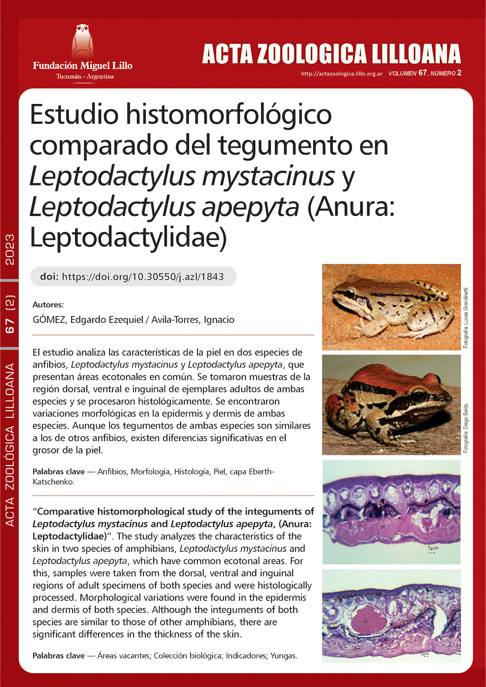
The study analyzes the characteristics of the skin in two species of amphibians, Leptodactylus mystacinus and Leptodactylus apepyta, which have common ecotonal areas. For this, samples were taken from the dorsal, ventral and inguinal regions of adult specimens of both species, from four specimens, two of each species, belonging to the Herpetological collection of the National Museum of Natural History of Paraguay (MNHNP) and were histologically processed. The skin micrometric data
were recorded and statistically analyzed with a confidence level of 95%, finding that the average skin thickness. Difference between species, in L. apepyta, the average thickness is 128.44 µm in the dorsal region, 81.36 µm in the ventral and 74.41 µm in the inguinal, while in L. mystacinus it is 139. 81 µm, 49.31 µm and 102.92 µm. in the same regions, respectively. Morphological variations were also found in the epidermis and dermis of both species, the statistical analyzes reveal a normal distribution of the data, concluding that, although the integuments of both species
are similar to those of other amphibians, there are significant differences in the thickness of the skin, with L. mystacinus being thicker. Furthermore, the presence of Eberth-Katschenko layers and glands with a differentiation in their maturation
is observed for both species.
Downloads
References
Azevedo, R. A., Carvalho, H. F., y de Brito-Gitirana, L. (2007). Hyaluronan in the epidermal and the dermal extracellular matrix: Its role in cutaneous hydric balance and integrity of anuran integument. Micron, 38(6), 607–610. https://doi.org/10.1016/j.micron.2006.09.008 DOI: https://doi.org/10.1016/j.micron.2006.09.008
Azevedo, R. A., Pelli, A. A., Ferreira-Pereira, A., de Jesus Santana, A. S., Felsemburgh, F., y de Brito-Gitirana, L. (2005). Structural aspects of the Eberth-Katschenko layer of Bufo ictericus integument: histochemical characterization and biochemical analysis of the cutaneous calcium (Amphibian, Bufonidae). Micron, 36(1), 61–65. https://doi.org/10.1016/j.micron.2004.06.004 DOI: https://doi.org/10.1016/j.micron.2004.06.004
Barthalmus, G. T. (1994). Biological roles of amphibian skin secretions. In H. Heatwole (Ed.), Amphibian biology (Vol. 1, pp. 382–410). Surrey Beatty & Sons.
Brunetti, A. E. (2014). Comunicación química de anuros: una perspectiva integral a partir de aspectos comportamentales, morfológicos y químicos en dos especies de Hypsiboas (Amphibia:Anura: Hylidae). Universidad de Buenos Aires.
Bueno, C., Navas, P., Aguirre, J. A., Aijon, J., y Lopez Campos, J. (1981). Skin Mucous Glands of Pleurodeles Waltii Mich. Histochemical and Ultrastructural Study. Archives de Biologie (Bruxelles), 92(1), 67–72.
Cei, J. M. (1980). Amphibians of Argentina (Vol. 2). Universitá degli studi di Firenze.
de Carvalho, C. B., Freitas, E. B. de, Faria, R. G., Batista, R. de C., Batista, C. de C., Coelho, W. A., y Bocchiglieri, A. (2008). História natural de Leptodactylus mystacinus e Leptodactylus fuscus (Anura: Leptodactylidae) no Cerrado do Brasil Central. Biota Neotropica, 8(3), 105–115. https://doi.org/10.1590/S1676-06032008000300010 DOI: https://doi.org/10.1590/S1676-06032008000300010
de Brito-Gitirana, L., y Azevedo, R. A. (2005). Morphology of Bufo ictericus integument (Amphibia, Bufonidae). Micron, 36(6), 532–538. https://doi.org/10.1016/j.micron.2005.03.013 DOI: https://doi.org/10.1016/j.micron.2005.03.013
Delfino, G., Brizzi, R., y Feri, L. (1995). Chemical skin defence in Bufo bufo: an ultra-structural study during ontogenesis. Zoologischer Anzeiger, 234(2), 101–112.
Denèfle, J. P., Zhu, Q. L., y Lechaire, J. P. (1993). Localization of fibronectin in the frog skin. Tissue and Cell, 25(1), 87–102. https://doi.org/10.1016/0040-8166(93)90066-T DOI: https://doi.org/10.1016/0040-8166(93)90066-T
Duellman, W. E., y Trueb, L. (1994). Biology of amphibians. The Johns Hopkins University Press Ltd. DOI: https://doi.org/10.56021/9780801847806
Elkan, E. (1968). Mucopolysaccharides in the anuran defence against desiccation. Journal of Zoology, 155(1), 19–53. https://doi.org/10.1111/j.1469-7998.1968.tb03028.x DOI: https://doi.org/10.1111/j.1469-7998.1968.tb03028.x
Farquhar, M. G., y Palade, G. E. (1965). CELL JUNCTIONS IN AMPHIBIAN SKIN. The Journal of Cell Biology, 26(1), 263–291. https://doi.org/10.1083/jcb.26.1.263 DOI: https://doi.org/10.1083/jcb.26.1.263
Felsemburgh, F. A., Carvalho-Silva, S. P., y de Brito-Gitirana, L. (2007). Morphological characterization of the anuran integument of the Proceratophrys and Odontophrynus genera (Amphibia, Anuran, Leptodactylidae). Micron, 38(5), 439–445. https://doi.org/10.1016/j.micron.2006.06.015 DOI: https://doi.org/10.1016/j.micron.2006.06.015
Felsemburgh, F. A., de Almeida, P. G., de Carvalho-Silva, S. P., & de Brito-Gitirana, L. (2009). Microscopical methods promote the understanding of the integument biology of Rhinella ornata. Micron, 40(2), 198–205. https://doi.org/10.1016/j.micron.2008.09.003 DOI: https://doi.org/10.1016/j.micron.2008.09.003
Felsemburgh, F. A., y de Brito-Gitirana, L. (2008). Avaliação morfológica do tegumento de fêmeas de Proceratophrys boiei. Espaço & Geografia, 11(1), 59–72.
Fox, H. (1986). Epidermis. In Biology of the Integument (pp. 78–110). Springer Berlin Heidelberg. https://doi.org/10.1007/978-3-662-00989-5_5 DOI: https://doi.org/10.1007/978-3-662-00989-5_5
Garcia, G. F., Cruz, P. I., y Mangione, S. (2011). Caracterización histomorfológica de la piel de especies de Leptodactylus del grupo fuscus (Anura: Leptodactylidae), destacando la capa de Eberth-Katschenko. Acta Zoológica Lilloana, 55(1), 33–43.
Gonçalves, V. F., y de Brito-Gitirana, L. (2008). Structure of the sexually dimorphic gland of Cycloramphus fuliginosus (Amphibia, Anura, Cycloramphidae). Micron, 39(1), 32–39. https://doi.org/10.1016/j.micron.2007.08.005 DOI: https://doi.org/10.1016/j.micron.2007.08.005
Greven, H., Zanger, K., y Schwinger, G. (1995). Mechanical properties of the skin ofXenopus laevis (Anura, Amphibia). Journal of Morphology, 224(1), 15–22. https://doi.org/10.1002/jmor.1052240103 DOI: https://doi.org/10.1002/jmor.1052240103
Heyer, M. M., Heyer, W. R., y de Sá, R. O. (2003). Leptodactylus mystacinus. In Catalogue of American Amphibians and Reptiles. University of Richmond. https://scholarship.richmond.edu/cgi/viewcontent.cgi?article=1072&context=biology-faculty-publications
Hoffman, J., y Katz, U. (1999). Elevated plasma osmotic concentration stimulates water absorption response in a toad. Journal of Experimental Zoology, 284(2), 168–173. https://doi.org/10.1002/(SICI)1097-010X(19990701)284:2<168::AID-JEZ6>3.0.CO;2-O DOI: https://doi.org/10.1002/(SICI)1097-010X(19990701)284:2<168::AID-JEZ6>3.0.CO;2-O
IUCN. (2022). The IUCN Red List of Threatened Species. Red List of Threatened Species. Version 2022-2. https://www.iucnredlist.org/
Katchburian, E., Antoniazzi, M. M., Jared, C., Faria, F. P., Souza Santos, H., y Freymüller, E. (2001). Mineralized dermal layer of the Brazilian tree-frog Corythomantis greeningi. Journal of Morphology, 248(1), 56–63. https://doi.org/10.1002/jmor.1020 DOI: https://doi.org/10.1002/jmor.1020
Langone, J. A., y de Sá, R. O. (2005). Re-description of the larval external morphology of two species of the Leptodactylus fuscus group (Anura, Leptodactylidae). Phyllomedusa: Journal of Herpetology, 4(1), 49. https://doi.org/10.11606/issn.2316-9079.v4i1p49-59 DOI: https://doi.org/10.11606/issn.2316-9079.v4i1p49-59
Lillywhite, H. B., Mittal, A. K., Garg, T. K., y Agrawal, N. (1997). Integumentary structure and its relationship to wiping behaviour in the common Indian tree frog, Polypedates maculatus. Journal of Zoology, 243(4), 675–687. https://doi.org/10.1111/j.1469-7998.1997.tb01969.x DOI: https://doi.org/10.1111/j.1469-7998.1997.tb01969.x
Mangione, S., Garcia, G., y Cardozo, O. M. (2011). The Eberth-Katschenko layer in three species of ceratophryines anurans (Anura: Ceratophrydae). Acta Zoologica, 92(1), 21–26. https://doi.org/10.1111/j.1463-6395.2009.00442.x DOI: https://doi.org/10.1111/j.1463-6395.2009.00442.x
Mangione, S., y Lavilla, E. O. (2004). Histología de la piel de la región lumbar de las especies sin glándula lumbar del género Pleurodema (Anura: Leptolactylidae). Acta Zoológica Lilloana, 48, 37–56.
Pianka, E. R. (1973). The Structure of Lizard Communities. Annual Review of Ecology and Systematics, 4(1), 53–74. https://doi.org/10.1146/annurev.es.04.110173.000413 DOI: https://doi.org/10.1146/annurev.es.04.110173.000413
Pough, F. H., Heiser, J. B., y Janis, C. (2008). A vida dos vertebrados (4th ed.). Atheneu.
Schneider, R. G., Cardozo, D. E., Brusquetti, F., Kolenc, F., Borteiro, C., Haddad, C., Basso, N. G., y Baldo, D. (2019). A new frog of the Leptodactylus fuscus species group (Anura: Leptodactylidae), endemic from the South American Gran Chaco. PeerJ, 7, e7869. https://doi.org/10.7717/peerj.7869 DOI: https://doi.org/10.7717/peerj.7869
Schoener, T. W. (1977). Competition and niche. In C. Gans & D. W. Tinkle (Eds.), Biology of the Reptilia (pp. 35–136). Academic Press.
Stiffler, D. F., Eskandari, S., y Dejbakhsh, S. (1997). Cutaneous transport in Ca2+ in the frog Rana pipiens: Significance and specificity. The Journal of Experimental Zoology, 277(5), 371–381. https://doi.org/10.1002/(SICI)1097-010X(19970401)277:5<371::AID-JEZ3>3.0.CO;2-M DOI: https://doi.org/10.1002/(SICI)1097-010X(19970401)277:5<371::AID-JEZ3>3.0.CO;2-M
Sullivan, P. A. (2000). Effects of Anion Substitution on Hydration Behavior and Water Uptake of the Red-spotted Toad, Bufo punctatus: is there an Anion Paradox in Amphibian Skin? Chemical Senses, 25(2), 167–172. https://doi.org/10.1093/chemse/25.2.167 DOI: https://doi.org/10.1093/chemse/25.2.167
Teixeira, S. A. M. V. (2014). Morfologia do tegumento de anfíbios anuros da Mata Atlântica e sua aplicação em estudos comportamentais. [Universidade Federal de Viçosa]. http://locus.ufv.br/handle/123456789/2372
Toledo, R. C., y Jared, C. (1993). The calcified dermal layer in anurans. Comparative Biochemistry and Physiology, 104A(3), 443–448. DOI: https://doi.org/10.1016/0300-9629(93)90444-9
Vitt, L. J., y Caldwell, J. P. (2013). Herpetology: an introductory biology of amphibians and reptiles (4th ed.). Academic Press. DOI: https://doi.org/10.1016/B978-0-12-386919-7.00002-2
Wanczinski, B. J., Barros, C. A. D. R., y Ferracioli, D. L. (2007). Hidratação do tegumento cutâneo. Revista Uningá, 12(1), 171–183.
Downloads
Published
How to Cite
Issue
Section
License
Copyright (c) 2023 Acta Zoológica Lilloana

This work is licensed under a Creative Commons Attribution-NonCommercial-NoDerivatives 4.0 International License.